In the intricate world of cell biology, the plasma membrane acts as a dynamic gatekeeper, orchestrating interactions that sustain life at the microscopic level. While textbooks offer schematic diagrams and static images, real stunning visuals reveal the membrane's complex heterogeneity and functional sophistication—providing insights that deepen our understanding far beyond traditional descriptions. This article peels back the curtain on five remarkable plasma membrane images that not only captivate the eye but also exemplify discovery and innovation in cellular imaging techniques.
Unveiling the Plasma Membrane: From Basic Models to Visual Masterpieces

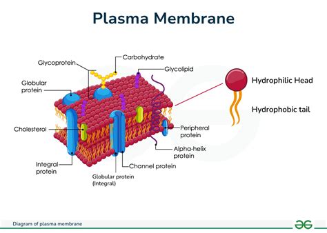
The plasma membrane, or cell membrane, is traditionally depicted as a fluid mosaic—a layered architecture of lipids, proteins, and carbohydrates. However, advances in microscopy and labeling technologies have transcended simplistic models, enabling scientists to capture living membranes in action with unprecedented clarity and detail. These cutting-edge images exemplify the culmination of multidisciplinary efforts in biochemistry, physics, and microscopy. They underline the importance of visual data in elucidating membrane architecture, dynamics, and function, while also presenting intriguing questions about cellular communication, signaling, and pathogen interactions.
Fluorescence Microscopy: Illuminating Lipid Rafts and Protein Clusters
One of the most awe-inspiring plasma membrane images springs from fluorescence microscopy, especially when combined with super-resolution techniques such as STED or PALM. These images reveal nano-scale domains—commonly referred to as lipid rafts—that serve as platform centers for signaling molecules. For example, a high-resolution fluorescence image might demonstrate a mosaic of vibrant, punctate clusters of cholesterol-rich regions interspersed with other membrane components. Such visualizations challenge the classical fluid mosaic paradigm by illustrating the membrane’s heterogeneity—and an active, organized landscape that adapts dynamically. They deepen our understanding of how membrane microdomains regulate processes like immune signaling, viral entry, and receptor localization.
Moving Beyond Light: Cryo-Electron Microscopy Revolutionizes Membrane Visualization

While fluorescence microscopy offers dynamic and live-cell compatible views, cryo-electron microscopy (cryo-EM) pushes resolution limits further, capturing detailed, near-atomic images of membrane components. Among the most stunning plasma membrane pictures obtained via cryo-EM are cross-sectional slices of vesicles or cell surfaces that showcase the bilayer’s bilinearity and embedded proteins in remarkable detail. These images often display the bilayer’s fluid mosaic architecture with specified lipid arrangements and integral protein conformations—truly revealing the membrane’s molecular sculpture. Such visuals have revolutionized our understanding of membrane protein complexes, channels, and transporters in native states, linking structural insights directly to functional understanding.
Atomic Models Derived from Cryo-EM Data: The Pinnacle of Structural Biology
Complementing cryo-EM images are detailed atomic models, sometimes shared as high-definition renders. These models act as precisely orchestrated reconstructions, allowing scientists to “walk through” the membrane landscape with true molecular fidelity. They portray complex assemblies like the voltage-gated sodium channel or G-protein-coupled receptors (GPCRs) in situ. The clarity of these 3D structures exemplifies scientific discipline’s mastery, providing critical insights into membrane protein mechanisms, drug targeting possibilities, and disease mutations—elements entirely missed in classical imaging.
| Relevant Category | Substantive Data |
|---|---|
| Resolution | Atomic models at ~2 Å resolution have been achieved, resolving individual amino acids within membrane proteins |
| Sample Preparation | Cryo-EM involves rapid vitrification preserving native structures without chemical fixation |
| Imaging Technology | State-of-the-art direct electron detectors improve signal-to-noise ratio and detail |

Correlative Light and Electron Microscopy (CLEM): Merging Modalities for Context-rich Views
The hybrid technique of CLEM combines the temporal and functional insights of light microscopy with the ultrastructural resolution of electron microscopy. Imagine an image series starting with fluorescence localization of a specific receptor on live cells, then correlating it with a high-resolution electron micrograph showing the underlying membrane architecture. Such images are visually stunning, often layered and color-enhanced to depict different molecules and structures simultaneously. The beauty of CLEM lies in its capacity to link function, dynamics, and structure seamlessly—serving as a powerful visual tool in membrane research, especially for understanding receptor clustering, lipid organization, or viral entry points.
Techniques Enabling Such Visuals
Critical to CLEM are innovations like fluorescent labeling compatible with electron microscopy contrast agents, sophisticated software for image registration, and advanced sample preservation methods. These concatenated images not only provide aesthetic appeal but also exceptional scientific value—allowing precise spatial correlation that leads to hypotheses on membrane behavior and organization at multiple scales, from nanometers to micrometers.
| Relevant Category | Substantive Data |
|---|---|
| Correlation Accuracy | Within 50 nm precision achieved in state-of-the-art systems |
| Applications | Studying viral fusion sites, receptor clustering, and membrane raft dynamics |
Conclusion: Visualizing the Pulse of Life at the Cell Membrane
The journey from basic membrane sketches to these mesmerizing images underscores a broader truth: visual data is crucial in translating the complexity of biological systems into comprehensible, actionable knowledge. The five plasma membrane pictures highlighted reflect not just technological prowess, but also a conceptual shift—a move toward seeing the cell surface as a vibrant, organized, and highly functional environment. As imaging technologies advance and analytical methods mature, we will continue to unlock deeper layers of understanding, each stunning image revealing new facets of the membrane’s role in life itself. For researchers and enthusiasts alike, these visuals are windows into the living, breathing entities that define biological existence—proof that science’s most beautiful revelations are often hidden in plain sight, waiting to be captured and understood.
Key Points
- High-resolution fluorescence microscopy exposes membrane microdomains like lipid rafts, vital for cell signaling.
- Cryo-electron microscopy provides near-atomic views of membrane protein complexes embedded within lipid bilayers.
- CLEM synergistically combines dynamic labeling with detailed ultrastructural context for comprehensive membrane studies.
- Advances in imaging technology continually push the boundaries of visualizing membrane heterogeneity and dynamics.
- These images serve as both scientific evidence and artistic representations of cellular life’s complexity.
What makes these plasma membrane pictures so significant for scientific research?
+They provide unparalleled visual insights into membrane architecture, organization, and dynamics, enabling precise understanding of cellular processes and facilitating targeted drug development.
How do cryo-electron microscopy images differ from fluorescence images of membranes?
+Cryo-EM offers atomic-scale resolution of membrane structures in a near-native state, while fluorescence imaging captures live dynamics and microdomain behavior at lower resolution but higher temporal detail.
What role does technology innovation play in producing these stunning images?
+Technological advancements like super-resolution microscopy, direct electron detectors, and correlative imaging are vital for achieving the clarity, resolution, and contextual depth seen in these membrane visuals.